Knee arthritis patient outcomes affected by significant hip weakness

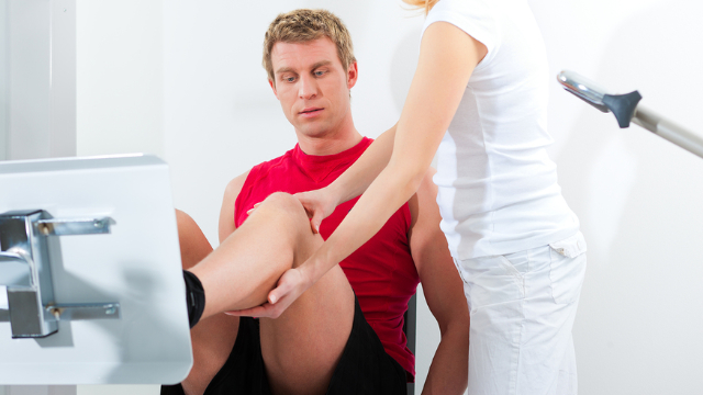
Understanding the role of hip strength can help improve treatments

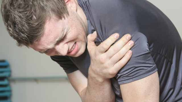
Osteoarthritis (OA) is a condition in which cartilage that normally protects a joint gradually wears down over the course of time, which leads to pain and other symptoms when the bones begin rubbing against one another. OA can develop in any joint, but is most common in the knee because it’s a joint that bears a great deal of weight. Knee OA is associated with high levels of pain and reduced function, and when the condition gets too bad, individuals often go on to have surgery to replace the knee joint. This shows why it’s so important to establish effective treatments for knee OA patients that will help them avoid surgery, but experts have pointed out that much more work needs to be done on the topic. One area of interest is the role of hip strength, as some have suggested that knee OA patients may have weak muscles surrounding their hips, and treatments can therefore target this weakness. With this in mind, a team of researchers performed a powerful pair of studies called a systematic review and meta-analysis to determine if patients with knee OA have deficits in their hip strength compared to healthy individuals.

Five medical databases searched for relevant studies

Investigators performed a search of five major medical databases for studies that evaluated the relationship between knee OA and the strength of patients’ hip muscles. They identified 102 studies that were screened in more detail, and five of these were accepted into the review, which contained data on 237 participants. The findings from each of these studies were then compared to one another, and their quality was assessed to determine how reliable they were.

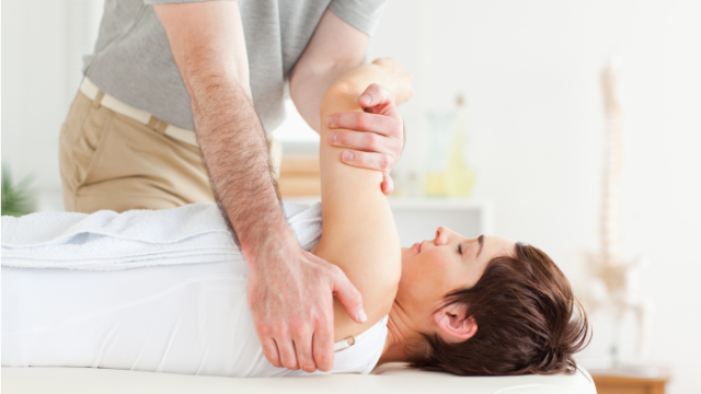
The weak hip muscles identified can be targeted in physical therapy programs

Overall, the quality of the studies reviewed was variable, but there was enough information to show that the hip muscles of knee OA patients were generally weaker than those in healthy individuals. In terms of isometric hip strength—the strength used if you were to push against an immovable object—there was moderate-quality evidence that knee OA patients have weaker hip abduction strength when compared with controls. (Abduction is moving the hip and the leg away from the center of the body.) When considering isokinetic hip strength—the strength that occurs when a muscle contracts and shortens at a constant speed—there was also moderate quality evidence that knee OA patients have weaker abduction/adduction and transverse internal/external rotation hip strength. Unfortunately, there were no studies that specifically evaluated hip strength as a risk factor for the development of knee OA. Nonetheless, this systematic review and meta-analysis show that patients with knee OA have deficits in their hip strength when compared to healthy individuals. Although more research is still needed on the topic, this could mean that improving hip muscle strength could lead to reduced pain levels and better hip function. Individuals with knee OA should therefore consider seeing a physical therapist for a comprehensive treatment program that will include hip-strengthening exercises to increase the chances of a successful outcome that does not involve surgery.

-As reported in the August ’16 issue of JOSPT

Patients treated with rehabilitation are less likely to use narcotics

The incidence of knee-related conditions has been increasing

Knee pain and knee osteoarthritis, sometimes referred to collectively as non-traumatic knee pain (NTKP), is very common in individuals aged 65 and older. Statistics have shown that the incidence of these conditions in older adults has increased by 65% over the past 20 years, with rates doubling in women and tripling in men. In addition, the rates for knee surgery have also increased over this period of time, which adds to the cost of managing these individuals. Several guidelines recommend rehabilitation such as exercise therapy to address pain and disability in patients with NTKP, which may also reduce the chances of these individuals using other dangerous treatment like opioids or needing to have surgery. Unfortunately, there is limited information on how many NTKP patients undergo rehabilitation, and it’s possible that not nearly enough are doing so. For this reason, a study was conducted to determine how many of these patients were receiving rehabilitation for their condition and examine how it affects their outcomes.

Data collected on extremely large group of patients

To conduct the study, researchers collected data on Medicare claims between 2009-2010. They looked for information on the utilization and cost of treatment for individuals over the age of 65 being treated for NTKP, which included 52,504 patient records. With this data, they examined which patients received rehabilitation services like physical therapy, how early this treatment started, what other treatments were delivered and how their outcomes differed depending on what treatment they received.

Only small percentage of patients are treated with rehabilitation services

Of the 52,504 NTKP patients examined, only 11% received any type of rehabilitation services for their condition at any time. Of the patients who were rehabilitated with physical therapy and other treatments, 52% began treatment within 15 days of their diagnosis, 27% started treatment within 16-120 days and 21% started treatment more than 120 days after being diagnosed. Further analysis showed that the participants who had early rehabilitation were 33% less likely to take narcotics like opioids, 50% less likely to receive invasive procedures like injections and 42% less likely to have surgery in the future than individuals who did not receive rehabilitation. Patients who didn’t receive rehabilitation until later, on the other hand, actually had higher odds of receiving these other types of interventions than those who did not receive any rehabilitation. These findings show that patients who are treated with physical therapy soon after being diagnosed with NTKP have better chances of improving without the use of dangerous narcotics or surgery, but only a small percentage are actually receiving this kind of treatment. It’s therefore important that doctors acknowledge the benefits of early rehabilitation for NTKP patients and refer them to physical therapy accordingly.

-As reported in the June ’17 issue of Physical Therapy

Improvements from PT for knee pain maintained by patients for 3 years

Long-term effects of treatment are questionable and have not been studied well

Patellofemoral pain syndrome (PFPS)-sometimes referred to as runner’s knee-is a painful condition that’s particularly common in physically active individuals. Patients with PFPS typically experience a dull ache behind or around the kneecap that gets worse with running or going up or down stairs. The initial treatment for PFPS is usually conservative (non-surgical) and may consist of a physical therapy program with various exercises that target the quadriceps muscles in the front of the thigh. While many studies have shown that this type of treatment is effective in the short term, long-term results are not as clearly understood. One reason for this is a lack of long-term studies that evaluate the potential benefits of conservative treatment. With this in mind, researchers conducted a follow-up study to investigate if improvements from a one-month physical therapy program in patients with PFPS were maintained three years later.

Participants are assessed at five different time points over three years

In the original study, 41 patients who had PFPS for at least one month were randomly assigned to one of four treatment programs: 1) quadriceps strengthening, 2) quadriceps stretching, 3) taping beneath the kneecap and 4) control, which did not receive any treatment. The total length of the intervention was one month, with the first week including specific exercises based on the groups participants were assigned to, and the second week consisting of a combination treatment of quadriceps strengthening, quadriceps stretching and kneecap taping. In the final two weeks, treatments were individualized to each patient and focused on aligning posture, correcting faulty movement patterns, improving knee and hip strength, stretching tight muscles and restoring flexibility. Patients were also given a home-exercise program with similar exercises to perform during these four weeks, and after completing this intervention, they were told to perform one quadriceps stretching and one quadriceps strengthening exercise daily for the next three years. All participants were assessed in seven categories at five different time points: before the intervention began, and then after one week, after two weeks, at the end of the intervention and three years later.

Majority of patients maintain their initial improvements and stick with exercise programs

Of the 41 original patients, 37 were available for the follow-up three years later. Overall, the measurements taken at this time were very similar to those taken at the completion of the intervention, which shows that patients maintained most of their improvements. In particular, all patients reported an improvement in knee pain scores when performing various physical activities, and 73% of them experienced no pain during the testing protocol. In addition, 28 of 34 patients (82%) were able to resume the sport that they had to stop because of PFPS, while three patients never played a sport in the first place. It was also found that 33 patients (89%) were continuing to do some of the exercises prescribed after completing the intervention, which probably helped improve their outcomes. The one measurement in which scores did decline was quadriceps strength, but this may simply be due to the fact that patients gained significant strength during the intervention. Nonetheless, these scores were still higher than those at the start of the study. These findings very clearly show that a physical therapy program for patients with PFPS leads to various improvements immediately after the intervention, and that these benefits appear to last for up to three years. Patients dealing with knee pain that may be related to PFPS are therefore encouraged to seek out treatment from physical therapist and to stick with their home-exercise program for the best chances of a successful long-term outcome.

-As reported in the May ’16 issue of the Clinical Journal of Sports Medicine

Course of exercise yields similar results as surgery for knee injury

Physical therapy may be utilized either instead of or after surgery

The meniscus is a crescent-shaped piece of cartilage between the thighbone (femur) and the shinbone (tibia), and its job is to stabilize the knee and absorb shock. Damage to the meniscus, which is called a meniscal lesion, can occur from an injury or due to gradual changes over time, which come with age. Patients with meniscal lesions who have knee pain and difficulty performing daily activities may be given different treatment recommendations depending on what type of doctor they see. Some patients are told to follow a wait-and-see policy, in which they rest for a period of time and see if their condition improves. Physical therapy may be utilized during this time. For patients with extreme pain and for those that do not improve, surgery may be recommended instead to repair the damage in the knee. In many cases, patients are referred to a course of physical therapy after surgery to help them rehabilitate. Treatment typically consists of a series of exercises to reduce pain and inflammation and help restore their normal range of motion. Although this is an approach that’s commonly recommended, it is not clear how effective exercise therapy is for these patients. Therefore, a powerful pair of studies called a systematic review and meta-analysis was conducted. The systematic review gathered all relevant research on exercise therapy and surgery for meniscal lesions, and the meta-analysis reviewed these all in detail to determine which approach is best for patients.

Nine medical databases searched for appropriate studies

To collect data for the review, researchers searched through nine major medical databases for studies on exercise therapy and surgery for meniscal lesions. Only randomized-controlled trials and controlled clinical trials—two types of powerful studies—that fit a set of criteria were accepted. This led to a total of 12 studies with data on 594 patients being accepted for the systematic review and meta-analysis. All studies were then analyzed and assessed for strength of evidence and risk of bias.

Exercise therapy found to have certain advantages over surgery

Overall, exercise therapy and surgery were found to lead to similar outcomes in knee pain, function, and performance for patients with meniscal lesions. Exercise therapy, however, was more effective for improving muscle strength than surgery after a short period of time. In addition, exercise therapy was found to be more effective than no treatment at all for muscle strength and performance in the short term. In the long term after surgery, exercise therapy was also more effective than no treatment on patients’ ability to function normally. Taken together, this suggests that patients who have surgery for meniscal lesions can benefit from exercise therapy after the procedure is performed. More research is needed to investigate this topic in greater detail, but it appears that exercise therapy can be considered an alternative option to surgery for treating meniscal lesions, and it may lead to similar overall results.

-As reported in the April ’16 issue of the Journal of Science in Medicine in Sport

ACL surgery patients similar outcomes as ones treated non-operatively

Patients’ expectations are not often met after surgery

Anterior cruciate ligament (ACL) tears are one of the most common injuries in athletes. These injuries are seen in a variety of sports, but athletes who participate in sports that involve lots of cutting movements-like basketball, soccer and football-are at a particularly high risk. Once an ACL tear occurs, the patient is usually presented with two options: treating it surgically with a procedure called an ACL reconstruction, or non-surgically through extensive physical therapy and rehabilitation. Many patients-especially athletes-decide to have an ACL reconstruction and go into the procedure with expectations that they will be able to return to their prior level of activity and avoid further injury. Unfortunately, these goals are not often met after surgery. In one study, only 65% of patients returned to their pre-injury level of sport, while 36% developed knee osteoarthritis within 10 years of surgery. But despite this information, many patients continue to have ACL surgery with lofty expectations that may not be met. To investigate this matter in more detail and better inform patients, a study was conducted to evaluate the differences in overall outcomes over five years in patients treated surgically or non-surgically for an ACL tear.

Majority of patients decide to have surgery

A total of 105 athletes between ages 14-55 years old, all of whom were involved in cutting and pivoting sports, were enrolled in this five-year study. These participants discussed whether they should have surgery or not with their surgeon and physical therapist, and 83 decided to have ACL reconstruction. The other 22 decided to have non-surgical treatment, which consisted of a physical therapy and rehabilitation program. After the program, patients in this group were instructed to follow a set of home exercises that focused on improving strength and muscular control, as well as helping them return to their former level of sports participation. Surgery was also followed up with a rehabilitation program that focused on bringing these patients back to their respective sport. All patients were assessed before the intervention began and then again five years later for a number of variables, including strength of the quadriceps muscles in the front of the thigh, a single-legged hop test and other patient-reported outcomes.

Positive outcomes occur in both groups

Five years after treatment, patients in both groups experienced positive outcomes, and the results between the two groups were found to be very similar. In particular, scores from the quadriceps strength and single-legged hop test were high and did not differ between the surgical and non-surgical groups. The same was true for the frequency of cutting and pivoting activities and the participation in pre-injury levels of these activities. Regarding overall activity levels, 61% of patients treated surgically and 50% of those treated non-surgically were able to engage in their pre-injury level of activity at five years. Patients in the non-surgical group reported scores that were slightly lower than the surgical group on quality of life, but scores were not different between groups in measures of knee symptoms or function. Finally, only 5% of patients treated non-surgically developed knee osteoarthritis by this time point, compared to 23% of patients treated surgically. Taken together, these results suggest that ACL reconstruction is not mandatory for all individuals who tear their ACL, and that positive outcomes are still very possible with non-surgical treatment, too. Therefore, if you recently tore your ACL and are currently trying to make a decision on whether or not to have surgery, keep this study in mind and have a thorough discussion with your surgeon and physical therapist. Every case is different, but it is possible that physical therapy alone may be sufficient for your condition and could help you achieve similar results to surgery without the associated risks, at a far lower cost.

-As reported in the June ’18 issue of The American Journal of Sports Medicine

Education program explaining pain is beneficial for patient outcomes

Patients generally want to learn more about their pain

Pain is necessary for our survival and something that we all experience to a certain degree from time to time. But dealing with pain on a regular or constant basis is not normal and requires appropriate treatment to address it. In addition to treating what’s called musculoskeletal pain—a term used to describe any pain that affects the bones, muscles or other connecting structures—education is another tool that may be helpful for improving the outcomes of patients. Studies have shown that people in pain are generally interested in learning more about what is causing their pain, and providing them with this education may reduce their pain and stress levels. There are several different types of educational strategies that focus on various systems of the body, but neuroscience education is believed to be most effective for patients dealing with continual pain. Pain neuroscience education (PNE) aims to explain the many processes at work involving the nervous system that causes pain, with the goal of making patients more aware of how to overcome it. In order to more develop a better understanding of the effectiveness of PNE, a powerful study called a systematic review was conducted. This type of study gathers all the most important literature on the topic and evaluates it in order to establish a conclusion.

13 high-quality studies accepted for review

Investigators searched through 11 major medical databases for studies that assessed the use of PNE for various musculoskeletal conditions. This search led to a total of 13 studies called randomized-controlled trials being accepted for the review. Randomized-controlled trials place participants in separate groups to receive different interventions, are they considered the highest-quality individual studies available. Each of these studies was evaluated in detail and then assessed for quality, which helps to establish how reliable its information is.

PNE reduces patients’ pain and improves their attitudes and behaviors

Results from the systematic review showed that PNE did, in fact, lead to several benefits for patients with musculoskeletal pain. In particular, it reduced patients’ disability and usage of healthcare and improved their pain ratings, knowledge of pain, as well as their attitudes and beliefs regarding pain. In addition, it was found that when PNE was combined with a physical intervention led by a physical therapist, patients experienced even better outcomes than when following the PNE alone. Finally, all the studies reviewed were rated as having good quality, which shows that their information was reliable. These findings show that PNE is effective for patients dealing with musculoskeletal pain since it can improve their attitudes and beliefs, and actually reduce their disability levels. More research is needed to explore the benefits of PNE further, but this type of education should be viewed as a helpful method for managing these patients.

-As reported in the July ’16 issue of Physiotherapy Theory and Practice

Thigh muscle exercises are beneficial for knee condition patients

Updated review of studies needed

Patellofemoral pain syndrome (PFPS) is one of the most commonly reported injuries in the field of sports medicine. The condition is most prevalent in runners and active individuals, and individuals who have it experience pain behind and around the kneecap that tends to get worse with squatting, prolonged sitting and stair activities. Treatment for PFPS typically consists of physical therapy, with a focus on exercises that strengthen the quadriceps muscles in the front of the thigh. This type of intervention is popularly used, but evidence to support its effectiveness is not very strong. The last review of studies on the topic was in 2003, and it only found little evidence that these exercises were effective for PFPS. For this reason, an updated review of the available literature-which is called a systematic review-was conducted, with the aim of determining the true effectiveness of quadriceps-strengthening exercises for PFPS patients.

Researchers perform a search of three databases for appropriate studies

To conduct the review, researchers performed a search of three major medical databases. They were specifically looking for high-quality studies called randomized-controlled trials (RCTs) that evaluated the effects of quadriceps-strengthening exercises for PFPS. RCTs randomly assign patients to various treatment groups and are considered the gold standard for determining whether an intervention works or not. This search led to seven RCTs being included: three RCTs on 204 participants investigated quadriceps-strengthening exercises alone, while five RCTs on 422 participants investigated quadriceps-strengthening exercises combined with other interventions. Once gathered, the researchers reviewed the findings of these RCTs and compared them to one another. The quality of all included studies was also assessed in order to gauge how reliable their results were.

Quadriceps-strengthening exercises alone and in combination reduce pain and improve function

On the whole, results were supportive of quadriceps-strengthening exercises for PFPS patients, both alone and when combined with other exercises. It was found that when they were performed alone, these exercises were more effective for reducing pain and improving function in patients when compared with advice and information alone. When combined with other interventions-such as hip-strengthening exercises, stretching, kneecap taping and home exercises-these exercises were also found to reduce pain for up to 12 months; however, they did not appear to improve function. This lack of improvement may be due to patients self-reporting their function scores, which has a margin for error. Nonetheless, the findings of this systematic review clearly show that quadriceps-strengthening exercises are beneficial to individuals with PFPS, whether used on their own or combined with other interventions. Based on this, patients currently dealing with pain that could be PFPS should think about seeing a physical therapist to receive an appropriate treatment program with these components to help them improve quickly and safely.

-As reported in the June ’14 issue of JOSPT

Pitching consecutive days in youth softball leads to pain & weakness

Popularity of softball is growing rapidly in the U.S.

Fast-pitch softball is one of the most popular and fastest growing sports among youth females, and the number of high school softball players in the U.S. is nearing that of baseball. But there is one major difference between the two sports: the mechanics of pitching a baseball and the impact it has on the body have been thoroughly studied, but research on the mechanics of the windmill pitch in softball is lacking. While it was once believed that the stress placed on the shoulder in softball is less than in baseball, recent studies have suggested that it may be more similar. The number of injuries in fast-pitch softball is also very high, but the details surrounding these injuries are poorly documented. But despite all these signs that the windmill pitching motion in softball is not as safe as previously thought, there are no pitching limitations in fast-pitch softball as there are in baseball. This means that some softball pitchers can pitch multiple games in a single day, up to three days in a row in some cases. Since research is lacking on the impact of pitching so frequently on these young pitchers’ arms, a study was conducted that investigated this effect on strength, fatigue and pain related to windmill pitching in youth softball.

Data collected on youth pitchers participating in 2-day and 3-day tournaments

For the study, researchers collected data on 17 female fast-pitch softball pitchers between the ages of 14-18 years who were participating in 2-day and 3-day weekend tournaments. Before pitching, information was gathered on these participants’ demographics and their history of shoulder injury or pain, as well as any other medical problems that might have had an effect on their ability to pitch. Then, at the start and end of each day of tournament play, these pitchers were assessed for fatigue, pain and strength of the shoulder and elbow in the dominant throwing arm. These values were compared at the end of each day and at the end of the tournament to determine if pitching had a negative effect on their symptoms.

Pain and fatigue are found to gradually increase over the course of the tournament

Of the 17 pitchers studied, eight participated in 3-day tournaments and six participated in 2-day tournaments. These athletes pitched in 1.5 games per day and threw 82 pitches per day, and there was a total exposure of three games pitched and 166 pitches thrown per tournament. Results showed that over the course of an average single day of tournament play, these pitchers had a significant increase in shoulder pain and fatigue, as well as a decrease in their strength. Over the course of the entire tournament, there was an even greater increase in pain and fatigue, as well as a greater decrease in their strength over this period of time. Finally, it was found that these pitchers did not recover to their baseline strength in many of the tested muscle groups by the day following pitching.

How pitch counts and physical therapy can help

These findings clearly show that windmill pitching on consecutive days leads to declines in shoulder and elbow strength that are not recovered the next day, as well as significant increases in pain and fatigue. These changes can lead to shoulder damage and increase the risk for injury in young softball players if they continue to pitch on consecutive days without regulations. This is why it has been suggested that implementing pitch counts similar to those used in baseball may address this potential problem and reduce the likelihood of injury in softball. Another possible solution to go along with pitch counts is to better prepare these young athletes for the demands of their sport with a conditioning training program provided by a physical therapist. Doing so can increase overall fitness levels and build up strength in these players so that their bodies are more capable of handling the impact of pitching regularly over the course of a softball season.

-As reported in the May ’17 issue of The American Journal of Sports Medicine

6 weeks of PT leads to improvements for athletes dislocated shoulder

One shoulder dislocation increases the risk of injuring it again in the future

The shoulder is the most frequently dislocated joint in the body, which is mainly due to the difference in size between the large end of the upper arm bone (humerus) and the small space it fits into (glenoid cavity). Shoulder dislocations usually occur after direct trauma or a fall onto an outstretched arm, and these injuries are common in sports like rugby, football, and hockey. Unfortunately, after a first dislocation, the shoulder can become unstable, which increases the chances of another dislocation occurring. This risk is particularly high in younger athletes and those involved in contact sports. Some studies have even suggested that the rate for regular shoulder dislocations is as high as 95% in young athletes who dislocate their shoulder once. These figures show why it’s so important to prevent these injuries from occurring in the first place and rehabilitating them properly when they occur. Physical therapy that focuses on strengthening the shoulder is one of the best ways to accomplish this, but it’s not clear which exercises are most effective towards these goals. Therefore, a study was conducted on the use of elastic bands and other strengthening exercises to improve joint strength in individuals diagnosed with a first-time shoulder dislocation.

Six weeks of therapy are divided into three stages

Researchers identified 12 physically active male athletes who had experienced a first-time shoulder dislocation who fit the necessary criteria to be included into the study. Two weeks after their injury, these participants began a six-week physical therapy rehabilitation program divided into three stages. The first stage consisted of a variety of strengthening exercises that were intended to build strength and control the pain and inflammation from the dislocation. These exercises were performed at a low intensity. The second stage focused on restoring a higher level of muscle strength and also increased flexibility, and the exercises were performed at a slightly higher intensity. The third and final stage focused on further improving endurance, strength and flexibility, and the intensity of the exercises increased even more. All stages of this physical therapy program included a variety elastic resistance bands for building strength in these muscles. Participants were evaluated before the program began and then again after two, four and six weeks for various measures of shoulder strength and flexibility.

Exercise program leads to injured shoulders becoming nearly as capable as uninjured shoulders

Results from the tests performed prior to the physical therapy rehabilitation program revealed that many patients had weakness in a number of their shoulder muscles as a result of pain, swelling and inflammation from the shoulder dislocation. After the six-week exercise program, however, these patients experienced considerable improvements of more than 90%. In fact, these improvements in strength and flexibility were so great that the values between the injured and uninjured shoulders were extremely similar in the final measurements. These findings suggest that a physical therapy rehabilitation program that includes elastic resistance bands and weight machines can be effective for improving strength and flexibility in patients who dislocate their shoulder. Following this type of treatment program after an initial dislocation may in effect reduce the likelihood of the patient experiencing another shoulder dislocation and needing surgery as a result. Therefore, while more research on the topic is needed, patients are strongly encouraged to visit a physical therapist after their first shoulder dislocation for appropriate treatment, as it may save them from additional shoulder injuries in the future.

-As reported in the June ’17 issue of the Journal of Exercise Rehabilitation

Mindset can have significant impact on patients with shoulder pain

Recovery rate is low for many of those with condition

Chronic shoulder pain-meaning it has lasted for at least three months-is a very common condition, with a prevalence of up to 67% in some populations. It is also a complex syndrome that features many unanswered questions, which is one reason the recovery rate from it is so low. Statistics show that about 60% of patients with chronic shoulder pain continue to deal with symptoms one year after first noticing it, and this highlights the need for better treatments to address the condition. Current treatments like physical therapy generally help patients in the initial stages of shoulder pain, but are less effective when it progresses into the chronic phase after several months. One reason for this is that psychological factors like depression, anxiety and fear of pain may actually impact the way in which the patient perceives pain and disability. The connection between psychological factors and chronic shoulder pain has been evaluated, but there is still a great deal that needs to be understood. For this reason, a powerful study called a systematic review was conducted to determine the extent that these factors can have on the perception of shoulder pain and what can be done to correct them.

Twenty-seven studies fit the necessary criteria

Researchers performed a search of five major medical databases for studies that investigated patients who had shoulder pain for at least three months. They were particularly interested in studies focusing on the psychological state of these patients and how it affected their pain and/or disability. This search process led to 128 studies being evaluated, and 27 of these fit the necessary criteria to be accepted into the review. Once collected, the findings from these studies were analyzed and compared to one another in order to establish a clearer idea of the connection between psychological factors and chronic shoulder pain.

Patients with positive attitudes about their condition have less pain and disability

The results clearly showed that patients’ attitudes and perceptions had a significant impact on their levels of pain and disability related to their condition. In particular, a strong relationship was identified between high levels of resiliency, confidence and positive expectations regarding recovery with low levels of pain intensity and disability. On the flip side, it was found that high levels of emotional distress, depressive symptoms, anxiety, concerns about surgery and fear of pain were associated with high levels of pain intensity and disability. The reason for these associations is not completely understood, but is believed to result from patients with negative beliefs perceiving their shoulder pain as a threat. In turn, this may lead them to stop using their shoulder and become less physically active, which begins a vicious cycle that only makes their condition and symptoms worse.

How physical therapists can educate and help these patients

These findings suggest that when treating shoulder pain, psychological factors must be taken into consideration and addressed in addition to physical symptoms. Physical therapists can help by identifying these negative factors in patients early on in treatment, and then work to educate and help patients overcome any barriers to success. In doing so, patients will understand their condition more clearly and increase their chances of experiencing a full recovery from shoulder pain. Patients with shoulder pain are therefore encouraged to consult a physical therapist at the earliest signs of shoulder pain for a full-scale evaluation and appropriate treatment program that takes all their physical and psychological symptoms into consideration.

-As reported in the April ’18 issue of BMJ Open